 by
by Types of Cardiac Pacing Leads
There are different types of leads used for cardiac pacing depending on the needs of the patient and where pacing is required in the heart. The main types include:
Venous Leads
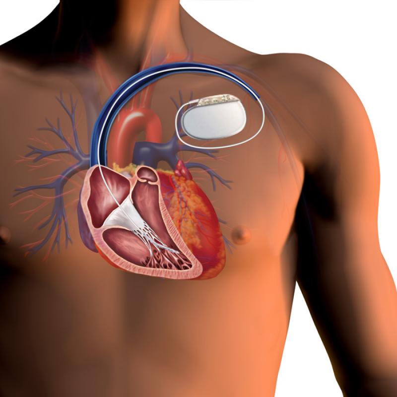
Venous leads are the most common type used as they can be inserted via a vein, usually the subclavian vein, and placed into the right-side chambers of the heart Cardiac Pacing Leads. There are three main types of venous leads:
– Right ventricular (RV) leads are temporary or permanent leads placed into the right ventricle. The RV lead is the most basic and commonly used lead for single-chamber pacing of the right ventricle.
– Right atrial (RA) leads are placed into the right atrium and used for single-chamber atrial pacing. RA leads help to pace the atrium when there is a problem with the SA node not firing properly.
– Dual-chamber leads have separate pacing electrodes that can be placed into both the right atrium and right ventricle. Dual leads allow both chambers to be paced sequentially for better synchronicity of heart contractions during dual-chamber pacing.
Coronary Sinus Leads
When pacing of the left chambers of the heart is required, coronary sinus leads are commonly used. These leads enter the coronary sinus, the large vein that drains blood from the heart muscle, which allows the electrodes to be positioned near the left atrium and ventricle for pacing. There are two main types:
– Left ventricular (LV) leads have distally placed electrodes that can be wedged into one of the coronary veins branching from the coronary sinus to pace the left ventricle.
– Bi-ventricular leads have additional electrodes that can pace both the right and left ventricles, enabling cardiac resynchronization therapy. This helps synchronize contractions between the right and left sides of the heart.
Epicardial Leads
Epicardial leads are placed directly onto the surface of the heart during open-heart surgery when transvenous leads cannot be used. The temporary or permanent epicardial leads are sutured onto the epicardium with the electrodes in contact with the heart muscle. This allows pacing of any chamber of the heart, but the procedure requires open-heart surgery.
Lead Functions and Components
All Cardiac Pacing Leads consist of similar basic components and functions:
– Pacing electrodes – The distally placed electrodes that come into direct contact with heart tissue. Multipolar electrodes can be programmed to different poles.
– Lead body – Flexible insulated wires or dual lumen tubes containing the coiled conductor wires running from the electrodes to the connector terminal.
– Connector pin – The proximal end connector that attaches into the pacemaker generator to enable electrical conduction from the generator circuitry to the heart tissue.
– Fixation mechanism – Helps secure the lead tip in position, such as passive tines or an active screw-in mechanism to prevent dislodgment over time.
Lead Implantation Procedure
The surgical implantation procedure of a transvenous cardiac pacing system involves:
– Venous access – A small incision is made and guiding sheaths/dilators are used to access and place leads in veins like subclavian, cephalic or jugular.
– Lead insertion – The lead is fed through the venous system using fluoroscopic guidance until the tip reaches the intended cardiac chamber.
– Lead positioning – Active or passive fixation is used to secure the lead tip in stable contact with the chamber wall.
– Pocket creation – A small pocket is made usually in the chest region subcutaneously for the pacemaker generator.
– Connector attachment – The lead proximal connector is attached to the generator before closing all incisions.
– Programming and testing – The pacemaker is programmed and lead function and sensing/pacing are tested.
This completes the system implantation with the leads anchored securely in the heart and pacemaker generator lying just under the skin in a hidden pocket.
Cardiac Pacing Lead Complications
As with any foreign medical device chronically implanted, there are risks of complications with cardiac pacing leads:
– Lead dislodgment – Leads can dislodge or migrate from their anchored positions over time.
– Twiddler’s syndrome – Wrapping of lead around its own axis from excessive lead manipulation.
– Insulation break or fracture – Fatigue or trauma may cause lead insulation or conductor wire breakage.
– Infection – Risk of pocket or systemic infection from bacteria traveling along the intravascular lead.
– Thrombosis – Blood clots can form on leads increasing risk of strokes.
– Pericardial effusion – Fluid buildup around the heart from lead perforation.
Therefore, implanted leads require regular follow-up to monitor for complications and ensure they are functioning properly for many years.
*Note:
1. Source: Coherent Market Insights, Public sources, Desk research
2. We have leveraged AI tools to mine information and compile it