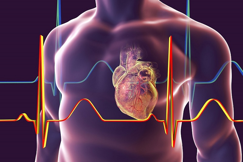
Electrodes Used During Pacemaker Placement
The most common electrodes inserted are endocardial pacing leads, which are thin, flexible wires inserted through a vein into the right ventricle or right atrium. Modern pacemaker leads are only a couple millimeters in diameter and constructed of careful insulated wiring able to withstand millions of bend cycles. Once positioned at the heart chamber’s interior wall, the lead tip contains a platinum exposed electrode designed to make stable contact with heart muscle tissue. Some newer lead designs have multiple electrode surfaces or stenosis to better anchor in position long term. The lead’s other end connectors to the pacemaker generator box implanted under the skin in the chest area. With electrode positioning verified, the generator is programmed to monitor the heart’s natural rhythm and provided backup pacing if the heartbeat becomes too slow.
Electrodes for Cardiac Mapping and Ablation
Advanced cardiac electrophysiology procedures use specialized Cardiology Electrodes with electrode tip designs to help map the heart’s electrical activity in detail. Multielectrode catheters like circumferential or linear array designs contain ring electrodes along the flexible catheter body or tip able to record signals from broad areas of heart tissue all at once. This provides high resolution mapping to identify abnormal conduction pathways or arrhythmias like atrial fibrillation. Catheters designed for cardiac ablation procedures have electrode tips capable of delivering radiofrequency energy pulses to create small scars in the heart muscle. This scar tissue interruption technique is used to treat conditions where abnormal electrical pathways cause irregular heart rhythms. By precisely guiding the catheter into position using electrode mapping data and fluoroscopy, doctors can effectively “burn away” problem areas generating arrhythmias. Follow up mapping verifies treatment success by ensuring the treated locations no longer conduct erratic impulses.
Implantable Loop Recorder Electrodes
For patients who experience infrequent episodes of unexplained symptoms like dizziness, an implantable loop recorder may be recommended. This small, encapsulated device contains miniature electrodes that continue recording the heart’s electrocardiogram for extended periods, from several days to years depending on battery life. During implantation via minor incision, the device is typically placed just under the skin above the left breast area. Its sensing electrodes lie just beneath the skin and are able to detect electrical signals transmitted from the heart muscle below. Whenever the patient feels symptoms, they use a wand device placed over the loop recorder to trigger memory of the preceding ECG data. Doctors can then download this information to determine if cardiac arrhythmias coincided with the events. Having continuous cardiac monitoring ability often allows implatable loop recorders to capture rare arrhythmic triggers that otherwise evade diagnosis with occasional Holter or event monitors.
Electrophysiology Study Recording Electrodes
One of the most in-depth cardiac evaluation procedures is the electrophysiology study Cardiology Electrodes , where multielectrode catheters are used to meticulously map the heart’s conduction pathways. At the start of the study, diagnostic catheter electrodes carefully record the heart’s baseline rhythms from the right atrium, coronary sinus vessel near the left atrium, and both ventricles. This involves threading multiple long, thin catheters with ring electrode arrays into the heart chambers through major blood vessels accessed via the groin or neck. If arrhythmias are induced in a controlled manner through catheter stimulation, precise recording of activation patterns helps identify the location of abnormal conduction or reentry circuits. Throughout rapid pacing maneuvers, extra beats, and even induced arrhythmia like atrial fibrillation, continuous recording with the multielectrode catheters allows electrophysiologists to visualize conduction in complex three-dimensional structures like the pulmonary veins and left atrium. This detailed mapping guides appropriate treatment whether with catheter ablation, implantable devices, or medications. The study results determine if reentrant or focal sources driving arrhythmias can be treated percutaneously or if other options are required.
Surface ECG Electrodes
Even the most basic cardiac diagnostic and monitoring tool involves electrode placement on the skin’s surface. Standard 12-lead electrocardiograms rely on 10 electrodes strategically placed on the extremities and chest wall to record the summated electric impulse spreading through the heart muscle from differing perspectives. This provides a global view of rate, rhythm, conduction, intervals and any signs of chamber enlargement, ischemia or injury that would be difficult to detect otherwise. During stress testing or Holter monitoring, additional limb or precordial leads track changes induced through activities or over extended periods. Surface electrodes also enable rhythmic monitoring during various medical procedures and recovery. Their noninvasive placement makes monitoring cardiac electrical activity relatively straightforward in many clinical settings. While not as detailed as intracardiac recordings, surface ECGs remain the cornerstone diagnostic modality for suggesting underlying cardiac anomalies, guiding treatment decisions and long-term tracking of conditions.
Note:
1. Source: Coherent Market Insights, Public sources, Desk research
2. We have leveraged AI tools to mine information and compile it


