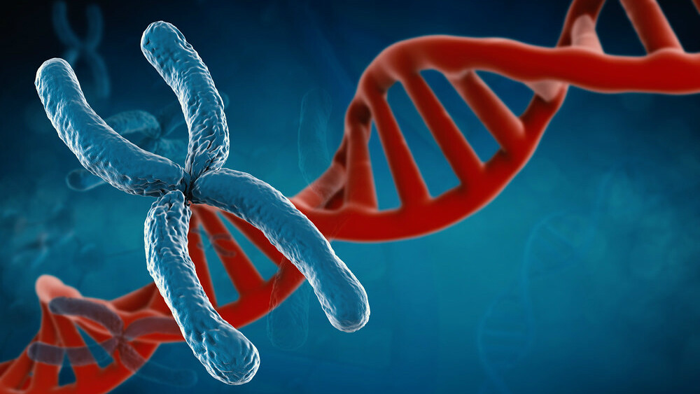
Molecular cytogenetics is a branch of genetics that combines microscopy-based chromosome analysis with molecular biology techniques such as DNA fluorescence in situ hybridization (FISH). The goal of molecular cytogenetics is to analyze chromosomes at a high resolution level in order to correlate disease phenotypes with underlying genetic changes or mutations. By employing both cytogenetic and molecular techniques, molecular cytogenetics has revolutionized our understanding of chromosome structure and genome organization.
Fluorescence In Situ Hybridization
One of the most widely used molecular Molecular Cytogenetic is fluorescence in situ hybridization or FISH. In a FISH experiment, fluorescent DNA probes are designed to complement specific DNA sequences of interest on chromosomes. These probes bind to only those sites on the chromosomes that are completely complementary to the probe’s sequence. By visualizing the fluorescent dots under a microscope, the position of the targeted DNA sequences can be mapped to specific chromosome bands. FISH has proven invaluable for detecting chromosomal abnormalities like deletions, additions, or translocations that are too small to be resolved by traditional cytogenetic analysis. It has enhanced our ability to diagnose diseases caused by such small defects.
Application in Cancer Research
Molecular cytogenetics techniques have extensive applications in cancer research. Most cancers are driven by genetic abnormalities that provide cells a growth advantage. FISH analysis has helped uncover recurring chromosomal rearrangements involved in different cancer types. Examples include the Philadelphia chromosome resulting from a translocation between chromosomes 9 and 22 in chronic myeloid leukemia (CML). Similarly, FISH probes have enabled the identification of gene fusions involved in diagnoses like Ewing sarcoma and Burkitt lymphoma. Molecular cytogenetics continues to refine our understanding of cancer genetics by revealing novel prognostics and targets for personalized therapy.
Prenatal Diagnosis Using Microarrays
In prenatal diagnostics, molecular cytogenetics has improved the detection of chromosomal abnormalities over traditional karyotyping methods. Microarrays employ DNA probes designed from across the human genome to hybridize with a sample of fetal DNA obtained from amniotic fluid or chorionic villus sampling (CVS). This fingerprinting technique allows for a higher resolution analysis than a karyotype and can identify copy number variations (CNVs) and other defects as small as five to ten megabases in size. Microarrays are now extensively used for prenatal diagnosis to screen for conditions like Down syndrome, Edwards syndrome, and Patau syndrome. They have enhanced our ability to counsel patients about risks of fetal anomalies pre-birth.
Visualization of Telomeres and Centromeres
Special FISH probes target repetitive DNA sequences located at telomeres and centromeres. Telomeres are found at the terminal ends of eukaryotic chromosomes and their visualization helps assess differences in telomere lengths linked to certain diseases or cancer. FISH probes specific for centromere repeat sequences allow direct examination of the number and structure of centromeres on each chromosome. Combined with staining for chromatid cohesion proteins, FISH has provided insights into sister chromatid interactions and the mechanism of meiosis. Molecular analysis of telomeres and centromeres helps uncover their roles in maintaining genome stability and driving genetic conditions.
Analysis of Structural Variations
Besides chromosome abnormalities, molecular cytogenetics techniques allow highly sensitive profiling of structural variations (SVs) in the human genome. These include deletions, duplications, inversions, and insertions. Traditional cytogenetics often misses SVs smaller than three to five megabases in size. However, molecular approaches employing microarrays coupled with FISH validation have enabled mapping SVs down to kilobase level resolutions. Genome-wide association studies frequently rely on molecular cytogenetics to precisely characterize SVs linked to complex traits and diseases like autoimmune disorders and neurodevelopmental conditions. Evolving technologies such as optical mapping and long read sequencing further aid comprehensive analysis of structural variations.
In summary, molecular cytogenetics leverages both optical and molecular techniques to study chromosomes at an unparalleled level of resolution. It has transformed our insights into causes of many genetic disorders and mechanisms of key cellular processes like mitosis and meiosis. Current areas of active research include single cell analyses, whole genome profiling of structural variants, and integration of multi-omics data. Molecular cytogenetics continues to unveil new genotype-phenotype correlations and power diagnostics, therapeutics and our basic understanding of human disease.
Note:
1. Source: Coherent Market Insights, Public sources, Desk research
2. We have leveraged AI tools to mine information and compile it


