
 by
by A groundbreaking method of visualizing brain activity using magnetic resonance imaging (MRI) was touted as a game-changer in neuroscience. However, researchers at MIT’s McGovern Institute have discovered that the purportedly direct detection of neural activity by this new MRI technique is actually a result of the imaging process itself, rather than neurons firing. The study, published in Science Advances by the lab of McGovern’s associate investigator Alan Jasanoff, reveals that the signals produced by this method do not accurately reflect neuronal activity.
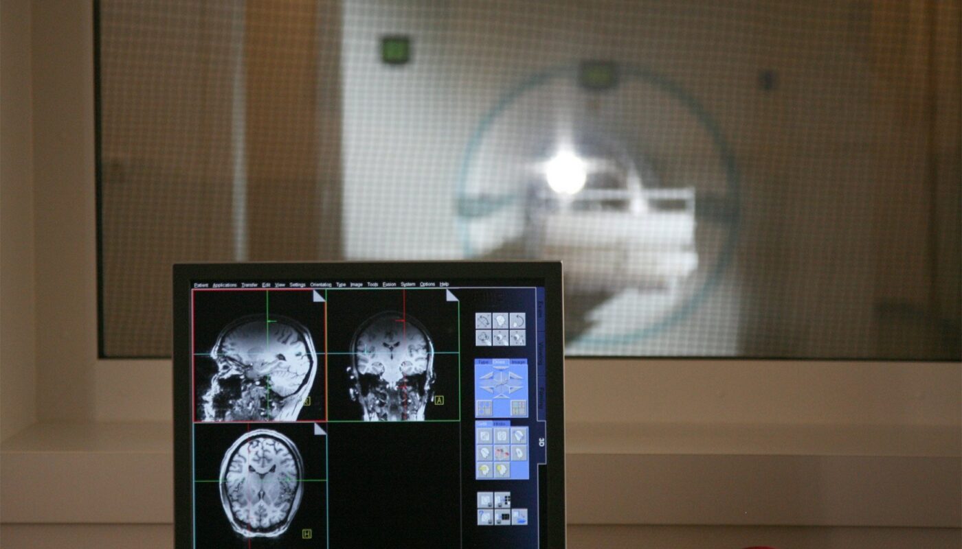
Traditionally, functional MRI methods rely on changes in blood flow to infer brain activity, providing a rough estimation of activated brain regions. While this approach offers insights into brain engagement, it lacks the precision to pinpoint neural activity at specific locations or capture the rapid communication between neurons.
The newly introduced MRI method, named DIANA (direct imaging of neuronal activity), promised real-time detection of neural signals with unprecedented speed and accuracy. Neuroscientists were intrigued by the potential of observing neuronal activity in the whole brain with millisecond precision, revolutionizing our understanding of brain function and disorders.
Initially intrigued by DIANA’s capabilities, Jasanoff’s team set out to replicate the results. Using the method to observe a rat’s sensory cortex while stimulating its paw, postdoctoral researcher Valerie Doan Phi Van initially observed what appeared to be neural activity correlated with the stimulus. However, further investigation revealed that the MRI signals persisted even when the paw stimulation was removed, indicating that the signals were not a true representation of neuronal responses.
Phi Van identified the source of these misleading signals in DIANA’s imaging process, specifically in the pulse program that synchronized the stimulus delivery with data acquisition. By modifying the trigger mechanism, she successfully eliminated the false signals associated with neural activity, confirming that the detected signals were artifacts of the imaging process.
The researchers also debunked the proposed cellular changes responsible for functional MRI signals suggested by DIANA’s developers, further emphasizing the method’s limitations in detecting true neuronal activity.
Jasanoff and Phi Van stress the importance of transparency in scientific research, urging caution when interpreting novel findings to prevent falling into methodological traps. While acknowledging the original study’s ambition, they highlight the necessity for rigorous validation and replication in advancing neuroimaging techniques.
By shedding light on the limitations of the DIANA method, the researchers aim to guide future developments in neuroimaging towards more reliable and accurate approaches, ultimately enhancing our understanding of the brain and its complexities. The journey to unraveling the mysteries of the brain continues, with each discovery refining our tools and techniques for probing its intricate mechanisms.
Note:
1. Source: Coherent Market Insights, Public sources, Desk research.
2. We have leveraged AI tools to mine information and compile it.